Microtubule-Mediated Granulosa Cell–Oocyte Communication and Its Role in Female Fertility
Thu, Jul 2, 2026-
Tags
New research reveals the role of Camsap3-organized microtubules in ovarian follicle communication and development
Researchers from Japan investigated how microtubules contribute to communication between granulosa cells and oocytes during ovarian follicle development. Focusing on the microtubule-stabilizing protein Camsap3, the team used knockout mouse models and advanced imaging techniques to examine the organization of transzonal projections. Their findings reveal an overlooked role for microtubules in female fertility and provide new insights into the cellular mechanisms that support follicle maturation and successful ovulation.
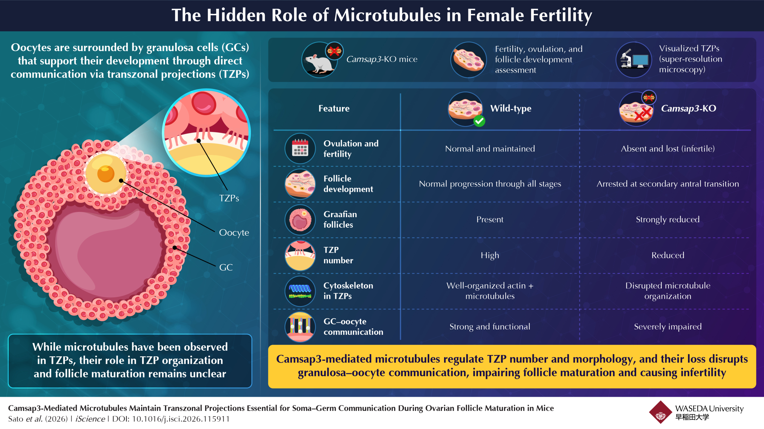
Image title: The Hidden Role of Microtubules in Female Fertility
Image caption: Disrupted TZP communication in Camsap3-KO mice leads to failed follicle maturation and infertility.
Image credit: Masamitsu Sato from Waseda University
License type: Original content
Usage restrictions: Cannot be reused without permission.
Female fertility depends on the successful growth and maturation of eggs (oocytes) within ovarian follicles. Within these follicles, the oocyte is surrounded by granulosa cells that supply nutrients, signaling molecules, and structural support essential for egg development. Communication between granulosa cells and the oocyte occurs through thin cellular extensions known as transzonal projections (TZPs), which cross the zona pellucida and physically connect the two cell types. Although TZPs were traditionally considered primarily actin-based structures, the role of microtubules within them remained poorly understood.
To better understand the contribution of microtubules to ovarian follicle development, a group of researchers from Japan, led by Professor Masamitsu Sato and Associate Professor Mika Toya from Waseda and Kyoto University, investigated the role of the microtubule-stabilizing protein Camsap3 in TZP organization and granulosa cell–oocyte communication. The research team included Dr. Akihiro Aikawa, Dr. Takao Tsurumaki, Dr. Erina Kuranaga from Kyoto University, and Dr. Junya Ito from Azabu University. The study was made available online on April 28, 2026, and was published in Volume 29, Issue 6 of the journal iScience on June 19, 2026 .
To investigate the function of Camsap3, the researchers generated Camsap3-knockout (KO) mice and compared them with wild-type mice. Fertility and ovulation were evaluated through natural mating experiments and hormone-induced superovulation assays. Ovarian follicles at different developmental stages were examined using histological analysis, while super-resolution microscopy and immunostaining techniques were employed to visualize TZPs, actin filaments, microtubules, and Camsap3 localization. Additional experiments assessed granulosa cell apoptosis and proliferation, and follicle reconstitution assays were conducted to determine the individual contributions of granulosa cells and oocytes to follicular development.
The study revealed several significant findings. Female Camsap3-KO mice were completely infertile and failed to ovulate despite maintaining normal estrous cycles, indicating that the defect was not hormonal in nature. Follicle development was disrupted during the transition from the secondary to the antral stage, resulting in increased follicular degeneration and a marked reduction in mature Graafian follicles. Most strikingly, super-resolution imaging demonstrated that more than 80% of TZPs contain both microtubules and actin, overturning the long-standing belief that TZPs are predominantly actin-based structures.
Explaining the significance of this discovery, Sato shares, “Super-resolution microscopy revealed that microtubules are present within TZPs that connect the oocyte and surrounding granulosa cells at a higher frequency than previously detected, highlighting an underestimated role for microtubules in oocyte-granulosa cell (GC) communication.” The loss of Camsap3 caused severe disorganization of microtubules within TZPs, reduced the number of TZPs, and led to the disappearance of specialized TNT-like TZPs that may facilitate the transport of large molecules and organelles such as mitochondria. As a result, communication between granulosa cells and the oocyte was significantly impaired, ultimately preventing proper follicle maturation and ovulation.
Further emphasizing the broader implications of the findings, Toya shared, “The study demonstrated that Camsap3 stabilizes microtubules within the TZP, revealing the molecular mechanism by which impaired communication between the oocyte and granulosa cells leads to infertility and follicular atresia.”
The discovery provides important insights into the cellular mechanisms underlying female fertility. By identifying microtubules as key structural and functional components of TZPs, the study expands current understanding of ovarian follicle biology and highlights Camsap3 as a critical regulator of reproductive function. These findings may also help identify new targets for infertility diagnosis and treatment while improving in vitro follicular culture systems used in reproductive research and assisted reproductive technologies.
In conclusion, this study uncovers a previously underestimated role of microtubules in ovarian follicle development and demonstrates that Camsap3-mediated microtubule organization is essential for maintaining granulosa cell–oocyte communication, successful ovulation, and female fertility.
Authors:Akihiro Aikawa1, Takao Tsurumaki1, Erina Kuranaga2, Junya Ito3,4, Mika Toya1,2, and Masamitsu Sato1,5,6
Affiliations:
1Laboratory of Cytoskeletal Logistics, Department of Life Science and Medical Bioscience, Graduate School of Advanced Science and Engineering, Waseda University, Japan
2Laboratory for Histogenetic Dynamics, Graduate School of Pharmaceutical Sciences, Kyoto University, Japan
3Laboratory of Animal Reproduction, School of Veterinary Medicine, Azabu University, Japan
4Graduate School of Veterinary Science, Azabu University, Japan
5Institute for Medical-Oriented Structural Biology, Waseda University, Japan
Title of original paper : Camsap3-mediated microtubules maintain transzonal projections essential for soma–germ communication during ovarian follicle maturation in mice
Journal:iScience
DOI:https://doi.org/10.1016/j.isci.2026.115911
About Professor Masamitsu Sato from Waseda University
Masamitsu Sato is a professor in the Department of Life Science and Medical Bioscience at Waseda University. He received his Ph.D. in Biophysics and Biochemistry from the University of Tokyo and has over 25 years of research experience in cell biology and molecular genetics. Professor Sato has authored more than 56 peer-reviewed publications. His research focuses on cytoskeleton dynamics, chromosome segregation, meiosis, non-coding RNA, and single-cell analysis using fission yeast models. His notable achievements include receiving the MEXT Young Scientists’ Prize and several competitive international research grants.