Taking an Innovative ACL Reconstruction Technique to the World
Tue, Nov 17, 2026-
Tags
Professor Kiyotaka Iwasaki of Waseda University’s Faculty of Science and Engineering is developing a “tissue-regenerative ligament” for anterior cruciate ligament (ACL) reconstruction and working toward its practical application. In August 2024, the Clinical Trial Notification was accepted by Japan’s Pharmaceuticals and Medical Devices Agency (PMDA), and the clinical trial began at Tokyo Women’s Medical University in November. How did Professor Iwasaki develop a technology with the potential to transform sports medicine? This article traces the story behind its development.
Challenges Facing Athletes with Ligament Injuries and the Limitations of Current Medical Care
Anterior cruciate ligament injuries occur in many sports. Approximately 19,000 ACL reconstruction procedures are performed each year in Japan and 175,000 in the United States. The high risk of reinjury after treatment remains a serious concern for athletes. Professor Kiyotaka Iwasaki of Waseda University’s Faculty of Science and Engineering is working to address this challenge.
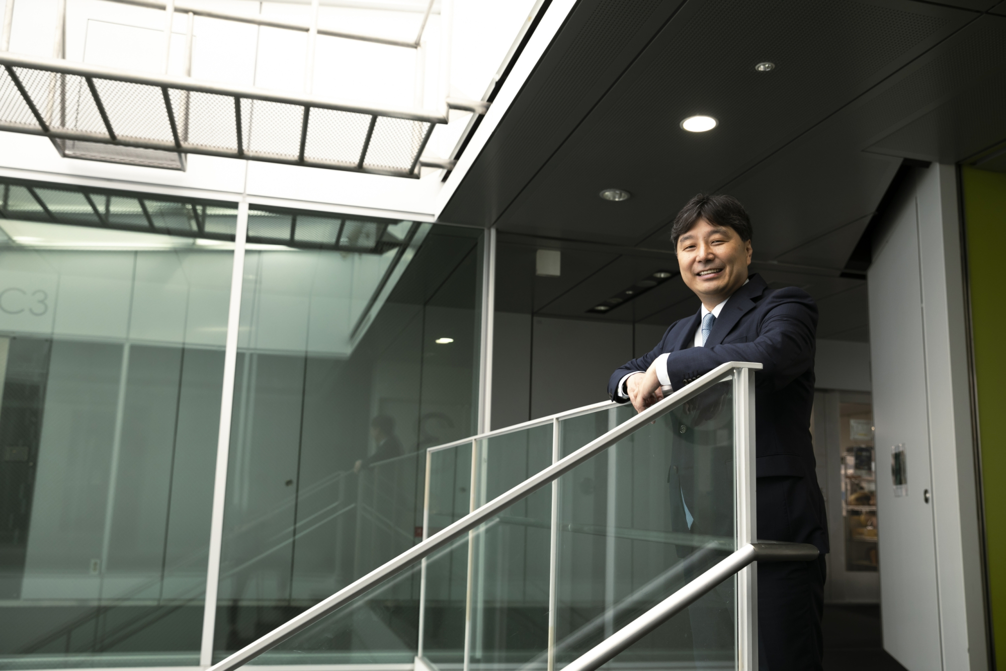
Professor Kiyotaka Iwasaki (Cooperative Major in Advanced Biomedical Sciences, Graduate School of Advanced Science and Engineering, Faculty of Science and Engineering). He is the founder and Chief Technology Advisor of CoreTissue BioEngineering, Inc.
“The anterior cruciate ligament in the knee is an important structure connecting the femur and tibia. When it is injured, athletes undergo reconstruction surgery in order to return to competition. However, with the current standard of care, it is difficult to eliminate the risk of reinjury after surgery. This reflects the limitations of conventional treatment.”
In ACL reconstruction, a substitute tissue known as a graft is secured in bone tunnels created in the femur and tibia. The graft is generally made from another tissue harvested from the patient’s own body, but Professor Iwasaki explains that this approach presents several challenges.
“The most commonly used graft materials are the hamstring tendon, located at the back of the thigh, and the patellar tendon harvested with small bone blocks attached. A thick tendon is needed to reduce the risk of postoperative graft failure, and a diameter of at least 8 mm is widely regarded as an important benchmark. However, hamstring tendons are relatively thin and vary from person to person. Even when folded into four strands, they are often less than 8 mm in diameter. Harvesting the hamstring tendon can also cause nerve injury or reduced muscle strength. With patellar tendon grafts, anterior knee pain may persist after surgery and knee-extension function may be impaired. In cases involving injuries to multiple ligaments, there may simply be insufficient tendon available for harvesting. Nevertheless, autograft reconstruction remains the approach used in almost all cases in Japan because there is currently no established alternative.”
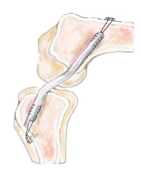
Location of the anterior cruciate ligament (left) and an illustration of the graft (right). A hamstring tendon graft is folded into four strands, but its average diameter is only 7.4 mm.
Developing a Thick, Strong Reconstruction Graft Using a World-First Decellularization Technology
Professor Iwasaki approached these challenges from an entirely new direction: using animal tissue rather than the patient’s own tissue. His team set out to create a reconstruction graft from bovine tendon.
“Mammals share broadly similar structural components, including collagen, and bovine tendons have sufficient thickness. However, transplanting them as they are would trigger inflammatory and other immune reactions, so the cells must first be removed from the bovine tissue. We therefore adopted a process known as decellularization.”
Professor Iwasaki’s decellularization technology removes cellular components from within the tissue without destroying its structural framework. Decellularization had existed since the late 1990s, but it was largely limited to thin membranes measuring no more than about 1 mm. Moreover, the solutions used to lyse cells could also damage the collagen structure. The technology therefore had to be advanced so that the graft could retain mechanical strength comparable to that of a human ligament.
“Our first idea was to apply microwaves, as in a microwave oven, to make water molecules vibrate. At 2.45 billion oscillations per second and a processing temperature below 37°C, the cell-membrane lysis solution can penetrate deep into thick tissue. We also introduced a system that circulates the solution through pulsatile flow and pressure. The inspiration came from the way the beating heart circulates blood throughout the body. We have obtained international patents for these two technologies, which reduce residual DNA to a level that meets safety criteria.”
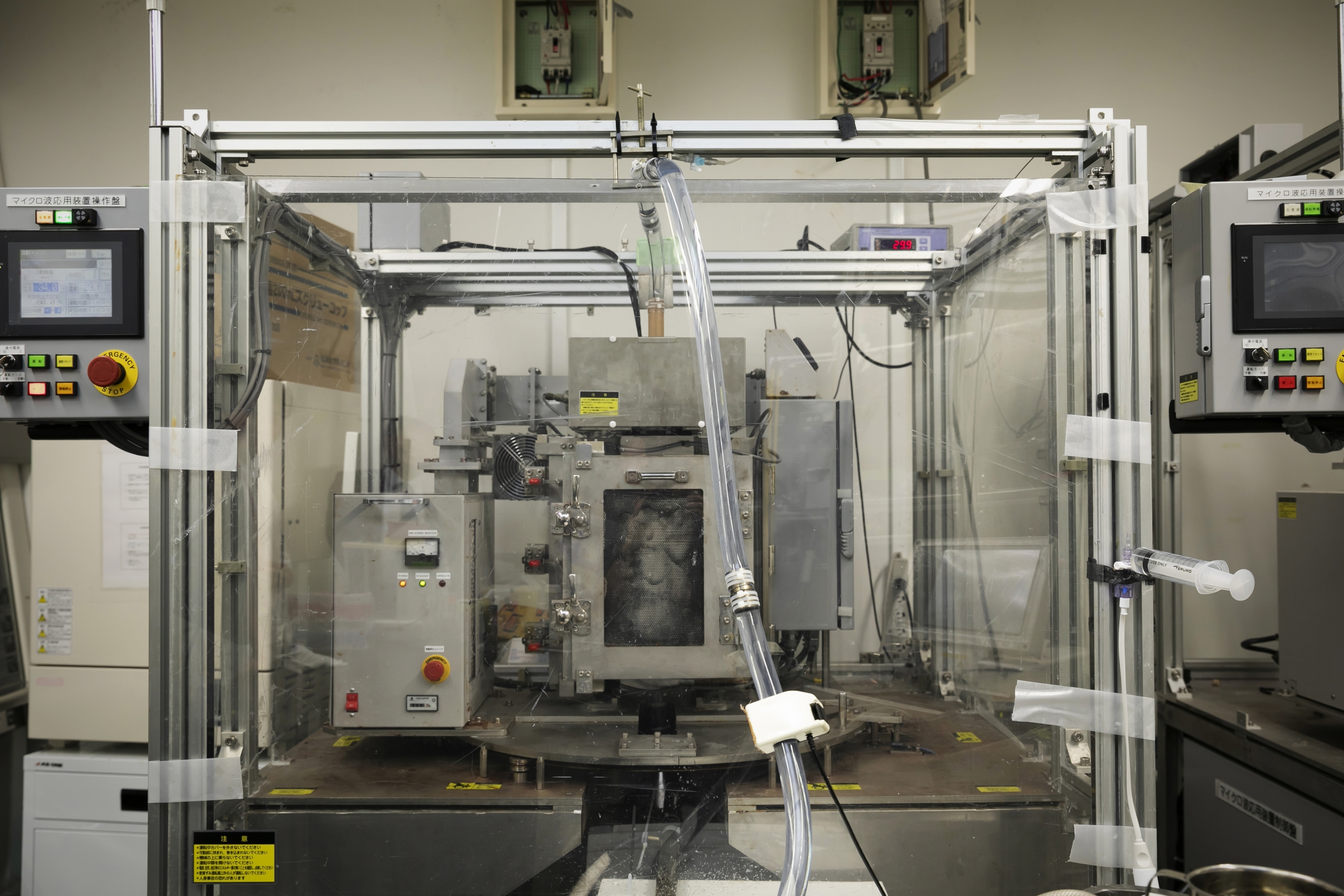
Equipment used in Professor Iwasaki’s decellularization process. While microwaves are applied to the tendon housed in the central chamber, a pulsatile pump delivers lysis solution through the tubing into the chamber, removing cellular components from the tissue.
Sterilization is also essential after the cells have been removed. Killing microorganisms without damaging the tissue is a major challenge in medical-device development. Gamma irradiation can degrade collagen, so the tissue must instead be freeze-dried and sterilized with ethylene oxide gas (EOG). The problem is that conventionally freeze-dried tissue does not readily rehydrate even when immersed in water. Professor Iwasaki needed to find a solution.
“The clue came from the sleeping chironomid, an organism that enters a dormant state during droughts in the African desert. We learned that it contains a distinctive sugar not found in most other organisms and decided to test it. By immersing the tissue in an appropriate concentration of sugar solution before freeze-drying, we were able to protect its mechanical strength and successfully restore it to a hydrated state after sterilization.”
The drying process. The team found that pretreatment in a sugar solution protects the tissue during both freeze-drying and EOG sterilization. This technology has also been patented internationally.
Commercializing a Self-Remodeling “Tissue-Regenerative Ligament” through a University Startup
The resulting decellularized tendon achieved mechanical strength comparable to that of human hamstring and patellar tendons. Human donor tissue is often too thin because of differences in body size and anatomy, but the new medical device combines the required thickness with high mechanical strength. Repeated animal studies conducted in preparation for practical application also demonstrated important biological properties.
“We performed ACL reconstruction in 56 sheep using either decellularized bovine tendon grafts or the sheep’s own tendons and compared the outcomes at approximately three months and one year. In the decellularized tendon group, the reconstructed tissue became firmly integrated with the bone. We also found that collagen density was higher at one year than at three months. This indicates that the sheep’s own cells had infiltrated the graft and begun regenerating functional tissue. Its defining advantage is excellent biocompatibility, something that cannot be achieved with synthetic materials.”
Firm bone integration and increased collagen density dramatically improve the stability of reconstructed tissue. By removing the cells while preserving the extracellular tissue architecture, the team created a “tissue-regenerative ligament” that can serve as a scaffold for the body’s own tissue remodeling.
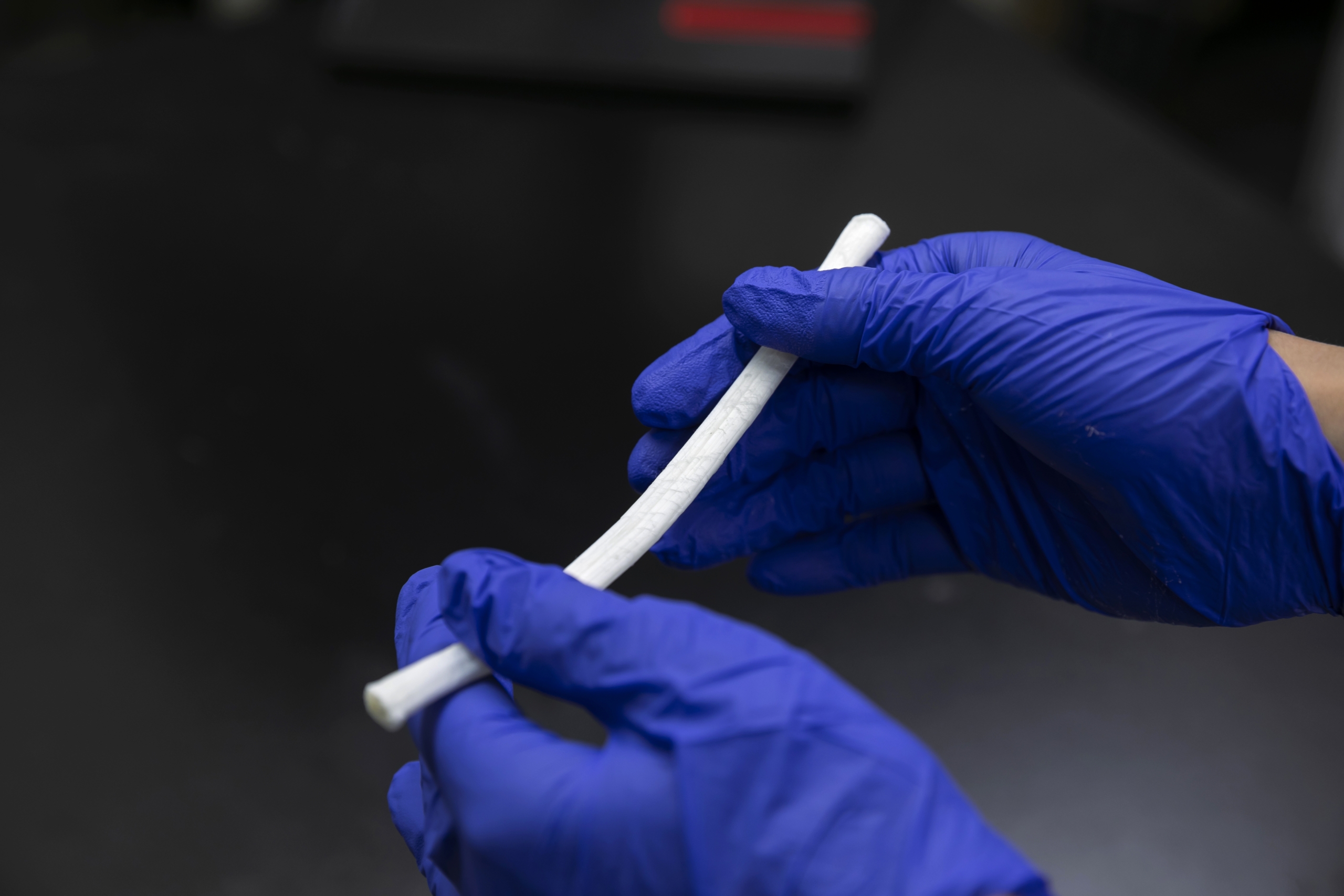
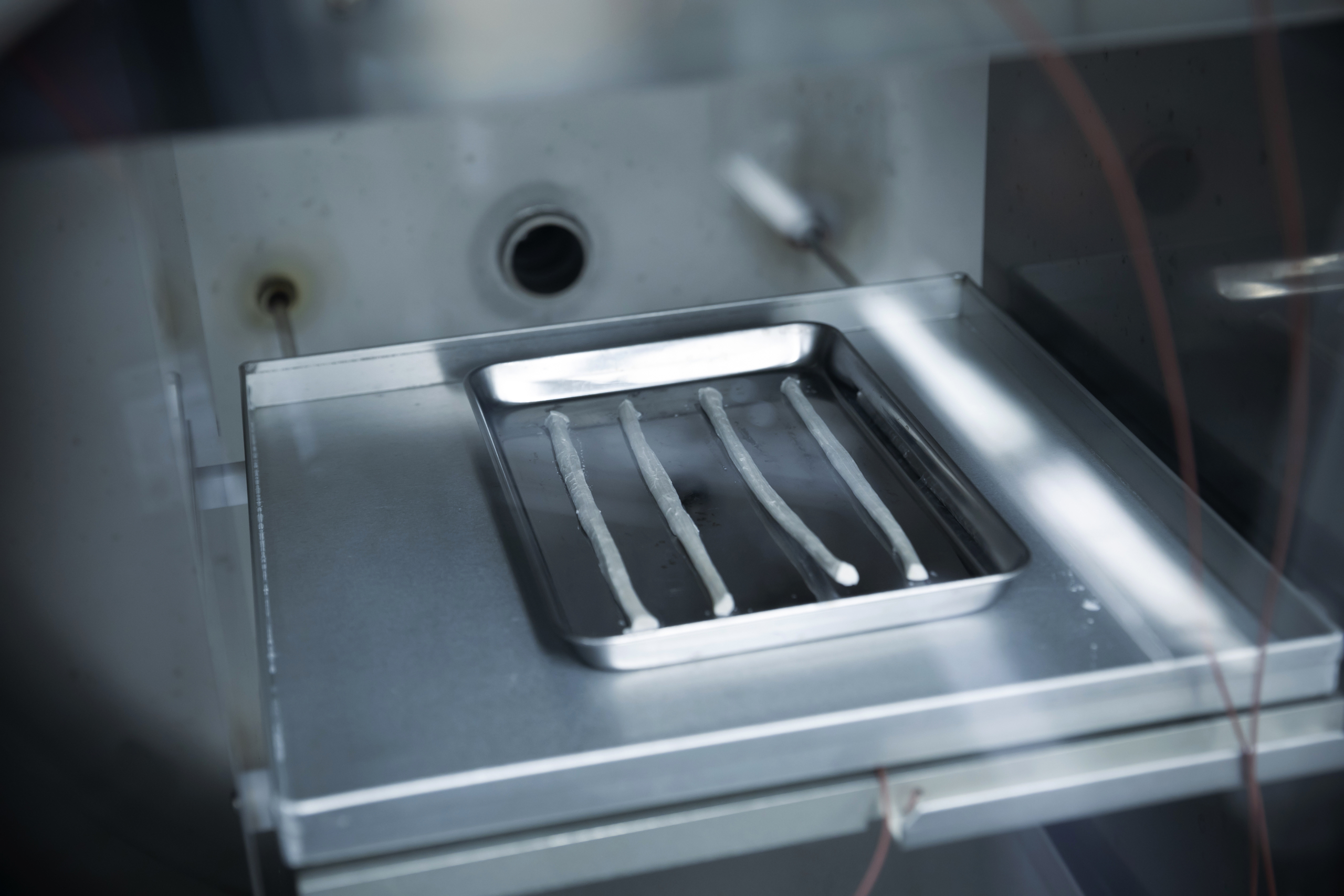
The decellularized tendon under development. It retains the elasticity and extensibility of biological tissue, while its clean white appearance gives little indication that it is animal-derived.
“If this technology can be applied in humans, the graft should gradually be replaced by the patient’s own ligament tissue inside the body. In a very literal sense, it will undergo ‘ligamentization’ and become integrated into the human body. To bring this world-first therapeutic device to patients, we established CoreTissue BioEngineering in 2016.”
Since the company’s establishment, CoreTissue BioEngineering has worked toward commercialization with support from the Japan Agency for Medical Research and Development (AMED), conducting preclinical research to evaluate safety and performance before application in humans. In August 2024, the Clinical Trial Notification submitted to the Pharmaceuticals and Medical Devices Agency (PMDA), under Japan’s Ministry of Health, Labour and Welfare, was accepted. The clinical trial began at Tokyo Women’s Medical University in November, and was scheduled to expand to six sites across Japan.
“After completing the clinical trial, the next step will be full-scale implementation. An estimated 800,000 patients worldwide require surgery for ACL injuries each year. We also believe that this technology could be applied to tendon and ligament injuries of the shoulder, elbow, and ankle. If we can steadily grow the business within this vast market, we may be able to bring meaningful innovation to sports medicine.”
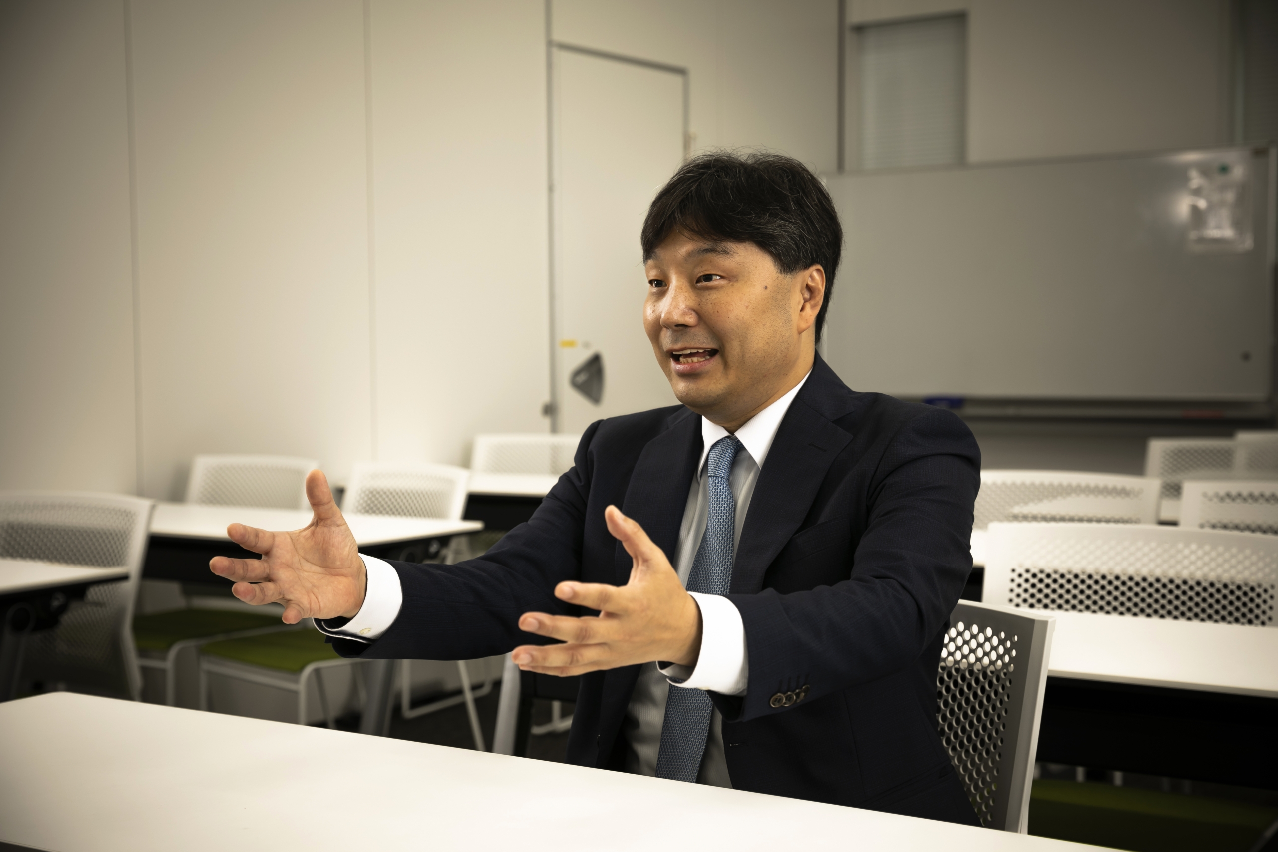
CoreTissue BioEngineering is currently establishing its production system and raising capital in preparation for implementation.
Contributing to Athletes Worldwide by Advancing Reconstructive Surgery
Professor Iwasaki is taking on the challenge of building a venture business around a world-first technology. What has motivated him to carry the project forward? We concluded by asking him about the commitment behind his research.
“From my undergraduate years in Waseda University’s School of Science and Engineering through the completion of my doctoral dissertation, I studied heart valves for use in artificial hearts. I then began researching decellularized tissues because I wanted to create therapeutic devices that would regenerate in the body and continue functioning for a lifetime. Later, researchers in sports medicine told me about the problems surrounding ligament reconstruction. Wanting to take on an unprecedented challenge, I began research on ligaments. I also enjoy sports myself, so I understand how important a stable ligament is. For athletes and everyone who participates in sports, a stable, functional knee without the burden of harvesting their own tissue, and the prospect of returning to activity even after repeated injury, would offer irreplaceable value.
“Nothing would make me happier than for this technology to relieve the concerns of as many people as possible and help them live fuller lives.”
Photo Credit: Waseda University Center for Advanced Biomedical Sciences (TWIns)