Now that its structure has been defined, scientists are a step closer to understanding the epigenetic gene regulation
Heterochromatin structure
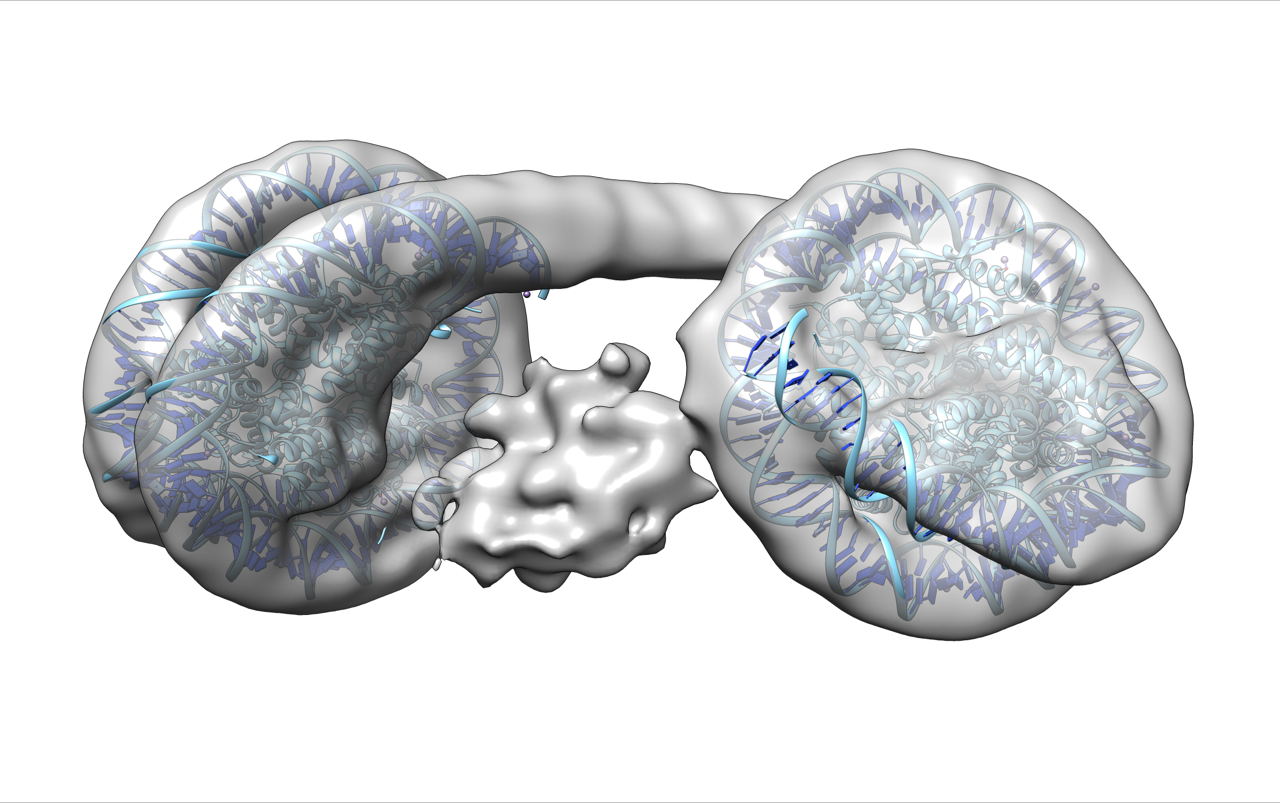
Heterochromatin is a chromatin which efficiently regulates genes due to its dense packing of the DNA. Though how the structural basis of heterochromatin is formed became clearer in recent years, the actual structure remained unknown since imaging it was difficult because of its flexibility and microscopic size.
According to a recent study published in Molecular Cell, scientists from Waseda University, Okinawa Institute of Science and Technology and the National Institute for Basic Biology became the first to successfully visualize the structure of heterochromatin.
“We discovered that heterochromatin looks something like a wireless headphone,” says Hitoshi Kurumizaka, professor of structural biology at Waseda University and leading scientist of this study. “Imaging heterochromatin became possible because of cryo-electron microscopy. Our study demonstrates Japan’s international competitiveness in structural biology research using this technique.”
The research group first reconstituted heterochromatin samples in vitro using a protein called heterochromatin protein 1 (HP1) and two nucleosomes linked together, which include histones mimicking a particular chemical modification (H3 lysine 9 tri-methylation). The samples were then purified for high-contrast imaging in cryo-electron microscopy, and from this image, the scientists found that heterochromatin is formed by HP1 binding with chromatin while bridging the nucleosomes positioned side by side.
Professor Kurumizaka points out that defining the heterochromatin structure could help better understand the mechanism of gene regulation and how certain kinds of diseases occur. “Damage to the heterochromatin structure is reported to increase chromatin abnormalities and cancer risks, and heterochromatin is closely associated with virus infections such as HIV. The results of this study could become valuable in developing treatment for such serious diseases.”
In the future, the scientists hope to study even more complex, higher order structures, such as an entire string of nucleosomes, using the method established in this study.
Reference
-
- Structural Basis of Heterochromatin Formation by Human HP1
- Published in Molecular Cell
- Authors:Shinichi Machida1, Yoshimasa Takizawa2, Masakazu Ishimaru1, Yukihiko Sugita2, Satoshi Sekine1, Jun-ichi Nakayama3, Matthias Wolf2, Hitoshi Kurumizaka1, 4
1 Laboratory of Structural Biology, Graduate School of Advanced Science and Engineering, Waseda University, 2-2 Wakamatsu-cho, Shinjuku-ku, Tokyo 162-8480, Japan
2 Molecular Cryo-Electron Microscopy Unit, Okinawa Institute of Science and Technology Graduate University, 1919-1 Tancha, Onna-son, Kunigami-gun, Okinawa 904-0495, Japan
3 National Institute for Basic Biology, 38 Nishigonaka, Myodaiji, Okazaki, Aichi 444-8585, Japan
4 Institute for Medical-Oriented Structural Biology, Waseda University, 2-2 Wakamatsu-cho, Shinjuku-ku, Tokyo 162-8480, Japan