MRI測定時の姿勢が大腿部筋量に及ぼす影響/Effects of postural conditions during magnetic resonance imaging on thigh muscle size

- Posted
- Mon, 06 Jan 2025
概要/Abstract
本研究では、男女若年ウェイトリフター20名および一般健常若年男女20名を対象にMRI撮像※1を行い、撮像時の姿勢が大腿部の筋量測定に及ぼす影響を調べました。撮像時の姿勢 (仰向け vs うつ伏せ) や大腿部とMRIの検査ベッドとの接触の有無によって大腿部の形状が大きく変わり、その影響を受け筋量も変化すること、特に男子ウェイトリフターのような筋量の大きい人ほど測定姿勢の影響が顕著であることが明らかとなりました。
In this study, we aimed to examine the effects of postural conditions during MRI on thigh muscle size. Twenty Olympic-style weightlifters and 20 untrained controls (10 men and 10 women in each group) underwent MRI measurements in the supine and prone positions, with the thigh in compressed and suspended conditions to determine the muscle volume of 15 thigh muscle groups/individual muscles. The results showed that the measurement position (supine vs. prone) and condition (compressed vs. suspended) substantially changed thigh morphology, and thereby muscle size. It has also been demonstrated that the effects of postural conditions vary across muscle groups and individual muscles. Furthermore, greater muscle volume changes were observed in male weightlifters, with the largest thigh musculature within the subgroups, suggesting muscle size dependence on postural-related muscle volume changes.
(1)これまでの研究で分かっていたこと/Research Background
MRIは骨格筋量を正確に測定する方法として研究・臨床現場で広く利用されています。特に大腿部には大腿四頭筋※2やハムストリングス※3といった日常生活動作やスポーツパフォーマンスにおいて重要な役割を果たす大筋群が存在し、これまで多くの研究が行われてきました。しかし、これまでの大腿部MRI撮像には、仰向けやうつ伏せ、大腿部をMRIの検査ベッドに直接置くか、検査ベッドとの接触による大腿部の圧迫を避けるためにクッションを使って浮かせるなど、多様な測定姿勢が用いられており、統一した撮像姿勢が使われていない、またこれらの姿勢の違いがどの程度大腿部の筋量に影響するかも不明という問題点がありました。
MRI is the gold standard for measuring skeletal muscle size; however, there are no standardized postural conditions during MRI measurements. In the case of thigh musculature, researchers have employed supine or prone positions, with the thigh directly placed on an MRI table or suspended between the hip and knee secured above the table. In either case, the thigh is compressed or sagged by gravity, which could affect muscle size.
(2)今回の研究で新たに実現しようとしたこと、明らかになったこと/Findings
本研究ではウェイトリフターと一般健常若年男女を対象に、仰向け・うつ伏せ × 大腿部の圧迫あり・なし、の4条件でMRI撮像を行い、大腿部の筋量を測定・比較しました (図1A)。その結果、MRI撮像時の姿勢および大腿部の圧迫有無によって大腿部の形状が大きく変わること (図1B)、またその影響を受け大腿部筋量も姿勢によって変化し、特に男子ウェイトリフターのような筋量の大きい人ほどその影響が顕著であることが明らかとなりました (図2)。
Twenty Olympic-style weightlifters and 20 untrained controls (10 men and 10 women in each group) underwent MRI measurements in the supine and prone positions, with the thigh in compressed and suspended conditions to determine the muscle volume of 15 thigh muscle groups/individual muscles. Regardless of measurement position, the total volume of thigh muscles decreased in the compressed conditions. Quadriceps and adductors decreased in muscle volume in the compressed conditions, while hamstrings increased. Male weightlifters, who had the largest thigh muscle volume, showed greater muscle volume changes in the quadriceps, hamstrings, and adductors than the other subgroups.
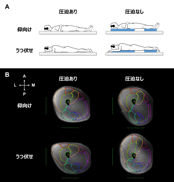
図1:本研究で用いた測定姿勢 (A) と各姿勢でのMRI大腿部横断画像の典型例 (B)。
比較のため、うつ伏せ条件での画像の向きは仰向けに合わせて反転させている。仰向け、うつ伏せそれぞれの姿勢において、大腿部は圧迫されると下部に位置する筋が平ら・横広に、圧迫なしでは縦長に変形することが見てとれる。
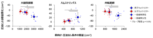
図2:大腿四頭筋、ハムストリングス、内転筋群※4の仰臥位・圧迫なし→圧迫ありの筋量変化。仰臥位において大腿部が圧迫されると、大腿四頭筋・内転筋群の筋量は減少し、意外にもハムストリングスの筋量は増加した。また、より筋量の大きい男子ウェイトリフターはより大きな筋量変化を示した。
(3)研究の波及効果や社会的影響/Research implications to the society
MRIによる大腿部筋量の測定は、アスリートの競技特性や、トレーニング・リハビリによる筋肥大効果、疾病や加齢に伴う筋委縮の程度を調べるために広く研究・臨床現場で行われてきました。本研究の結果から、これまでの測定姿勢が無根拠に設定されていた状況を見直し、大腿部MRI撮像時の測定姿勢を目的に応じて標準化することが必要になってくるのではないかと考えています。
Measurement of the thigh musculature and its adaptive changes has been extensively studied using MRI in research and clinical settings to investigate sport-specific muscle characteristics, muscle hypertrophy with mechanical loading, and muscle atrophy associated with unloading and aging. Based on the present results, it will be necessary to review the current situation where some have employed supine or prone positions and the thigh either placed on the examination table or suspended to avoid contact without any references. Our results suggest the need to standardize the optimal postural conditions for intra- and interindividual comparisons of thigh muscle sizes.
(4)今後の課題/Future issues
本研究では健常若年男女の大腿筋のみを対象としました。そのため、例えば中・高齢者やサルコペニアなどで極端に筋量が委縮しているケースに対しても一般化できるかどうかはわかっていません。また、撮像姿勢の違いにより生じる筋の変形・筋量の変化は、恐らく上肢や体幹、殿部、下腿など他の部位でも起こり得ますが、その程度は不明のままです。
We only examined the thigh muscles of healthy young individuals. Thus, it remains unclear whether our findings can be generalized to different muscles (e.g., upper limb, trunk, hip, and lower leg muscles) as well as other populations (e.g., elderly with sarcopenia and obesity).
(5)研究者のコメント/Researcher’s Comment
塩谷彦人:共同研究者としてMRI測定に携わる中で、根拠なく姿勢条件を設定している先行研究が多いことが気になり、「それでいいのか?」という疑問から着想に至りました。方法論的な内容ですが、筋量を正確に測る上で見逃せない重要なポイントを示した研究と考えています。また本研究は、本学部(早稲田大学スポーツ科学部)に設置されたMRI施設を活用し、早稲田大学ウェイトリフティング部員の皆さんに被験者としてご協力いただくことで実現できた、本学部ならではの成果と言えます。このような形で論文発表に至ることができ、大変嬉しく思っております。
川上泰雄:すさまじく大きな力を発揮する下肢の骨格筋は量も多く、水分も多く含むために、安静時は柔らかく、重力方向に垂れ下がります。このことは、骨格筋の研究者にすらあまり認識されていません。この点に注目し、姿勢による体積の変化にまで踏み込んだ分析をした本研究の意義は今後高まっていくことが期待されます。
(6)用語解説/Terminology
※1 MRI:磁気共鳴画像法 (Magnetic Resonance Imagingの略)。装置から発生する電磁場と体内に含まれる水分子の信号を利用して、人体の構造や情報を非侵襲的に画像化する医用画像手法。
※2 大腿四頭筋:大腿部前面に位置する筋肉の総称。人体で最も体積が大きく、股関節の屈曲や膝関節の伸展に作用する。
※3 ハムストリングス:大腿部後面に位置する筋肉の総称。股関節の伸展や膝関節の屈曲に作用する。
※4 内転筋群:大腿部内側に位置する筋肉の総称。主に股関節の内転に作用する。
(7)論文情報/Journal Information
雑誌名/Journal:Scandinavian Journal of Medicine & Science in Sports
論文名/Title:Effects of Postural Conditions During Magnetic Resonance Imaging on Thigh Muscle Size
執筆者名・所属機関名/Authors and Affiliated Organisation:塩谷彦人(早稲田大学)、西野優作(早稲田大学 スポーツ科学部23年度卒業生)、一瀬星空(立命館大学)、川上泰雄(早稲田大学)
Publishment Date(Local Time):14 November 2024
Publishment Date(Japan Time):15 November 2024
URL:https://onlinelibrary.wiley.com/doi/10.1111/sms.14760
DOI:10.1111/sms.14760